The Effect of Class I Anti-Arrhythmics

I’ve been asked quite a bit about the effect of class I anti-arrhythmic drugs.  The three subclasses of these drugs have different effects but they share one thing in common:  they block Na channels.

Na channels in ventricular and atrial cells much are like the Na channels in nerve and skeletal muscle.  They media rapid depolarization once threshold is attained.  Decreasing the number of available Na channels – in this case by blocking them – decreases action potential conduction velocity just like it did in these other tissues when we studied them previously.

Retrograde Conduction
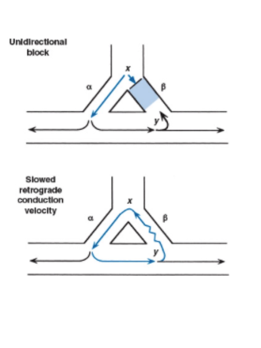
Consider the situation illustrated above.  The unidirectional block is in the beta branch of the pathway (top figure).  The action potential travels down the alpha branch (which is normal) and back up the beta branch (bottom figure).  Note that though conduction is still allowed up the beta branch in a retrograde fashion, these cells are still abnormal and conduction may be slowed (bottom figure, squiggly line).  If the action potential goes up the beta branch and reaches the alpha branch after the alpha branch has exited its refractory period, a re-entrant arrhythmia results.

With all of the class I anti-arrhythmic subclasses, the hope is that you will slow the conduction down in the already diseased beta branch enough to where it just completely stops and never gets back up to propagate back down the alpha branch.  Some of the subclasses are more effective than others in this regard.  And, of course, they all have different effects in addition to this.  But they all do the above to at least some extent.

Posted in Cardiovascular Block, Frequently Asked Questions | Leave a comment

Why Do the Atria Beat at a Faster Rate with Heart block?

I’m getting this one a lot:

I have a question regarding #28 on one of your old exams: “If the AV node is destroyed then it is expected that…”
I understand that “b) the ventricles will beat at a slower rate than normal” because their pace will be set by the purkinje fibers. But would “d) the atria will beat at the normal rate” not also be true because their contraction is dictated by the SA node and depolarization occurs independent and before the AV node?

Third degree heart block is, of course, what you get when the AV node can’t conduct action potentials to the ventricles.

Action Potential Propagation around the Heart

Propagation of the action potential around the heart. The numbers are the time in seconds that it takes for the action potential to reach each region as it propagates away from the SA node.

Physiologically, the SA node, the AV node and some Purkinje fibers all will spontaneously depolarize. In this case, the action potentials aren’t getting to the ventricle. Without action potentials from the the SA node, the AV node takes over first and if that can’t take over due to disease, the Purkinje fibers do. The SA node set the pace when healthy because it depolarizeS fastest during phase 4. The action potential propagates away and stimulates the AV node and Purkinje fibers before they have has a chance to spontaneously throw an action potential on their own.

Therefore you may conclude that the SA node sets the pace at a higher rate than the AV node or Purkinje fibers would.  If the SA node action potentials aren’t making it through, the AV node sets the pace at its own rate, which is slower or, if that’s not possible, the Purkinje fibers set an even slower rate.

Therefore:

Heart Block Concept Map 2015-11-19

Posted in Cardiovascular Block, Frequently Asked Questions | Leave a comment

The Effect of Exercise on Mean Arterial Pressure

I’ve been asked several times about question 27 on page 46 of the week 4 course notes.  The question asks about the steady state MAP during vigorous exercise.  The answer is that MAP is increased.

Exercise
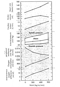
The problem is that, as most of you will recall, in the lecture I did in the Human Simulation Laboratory I said that the MAP wouldn’t change or might decline a little bit with an initial drop followed by a reflexive increase that wouldn’t quite get back up to the starting point.

In fact, if you look at the real data in the figure in that lecture, the MAP increases just slightly in this figure.  I don’t emphasize this in class because its due to a little understood central mechanism and the effect is very slight.


Posted in Cardiovascular Block, Frequently Asked Questions | Leave a comment

The Effect of Class IB Anti-Arrhythmics on Action Potential Propagation

I’ve been asked several times about this because I put a question on the practice exam related to it.

In retrospect, I wish that I hadn’t used the class IB as the example on this question and I will change it next year.  However, for the record, all of the class I anti-arrhythmics block Na channels and blocking Na channels slows the action potential propagation.  I admit that the effect is nowhere near as strong with the IB as it is in the IC and IA because the IB are open channel blockers that aren’t as effective during the rapid upstroke.  Nevertheless, it will always happen to some extent and it is an effect which you may see particularly in diseased tissue.

Posted in Cardiovascular Block, Frequently Asked Questions | Leave a comment

Where Does Ventricular Systole Start and End?

I’ve been asked this question several times now and it’s not unreasonable.  I have some thoughts:

  1. This is kind of a grey area and it depends upon the context when asking the question.  “Ventricular systole” when you are a physician doing a physical exam might be defined differently then when you are a scientist looking at the contractile properties of the cardiac muscle or an echo cardiologist watching the ventricle as it contracts and relaxes.
  2. Generally speaking everyone will agree that ventricular systole starts when the ventricular muscle starts to contract.  Some may prefer to define it as when you see the QRS complex on an ECG, as technically that is when contraction begins to be initiated. If you are listening to heart sounds, you will likely define ventricular systole as beginning with the first heart sound.  This is certainly almost simultaneous with when significant contraction begins to take place and pressure begins to build in the ventricle.
  3. Generally speaking every will agree that ventricular systole ends when the cardiac muscle stops contracting and begins to relax.  If you are looking at the ECG, that’s when you see the T wave.  If you are listening to heart sounds you will likely define ventricular systole as the period between S1 and S2 despite the fact that the T wave precedes S2 on the Wiggers diagram.  You will also likely define a “holosystolic murmur” as one that you hear roughly between S1 and S2, e.g. when you would hear an stenotic aortic valve or when you would hear an insufficient mitral valve.

    Wiggers

    Wigger’s Diagram

  4. I get the impression that students think that we’re going to ask you “What is the period of systole defined by” and treat is as if it’s a black and white line on the exam  and then scream, “Gotcha!” when you get it wrong because you chose to end it at the T wave when I chose to end it at S2.  If that’s what you’re worried about, don’t.  I’m not in the business of testing that kind of trivia and it should be clear what I’m looking for should I ask a question of this type.  On the other hand, if I say that a patient has a holosystolic murmur, you should generally know what that means and draw conclusions based upon the physiology of the situation.


Posted in Cardiovascular Block | Leave a comment

How Can the Conductance be Greater For K+ than for Na+ But the Currents Be Equal and Opposite?

I’m getting this one a lot.  Its #21 in the on exam questions after the Synaptic Transmission lecture but #7 after the resting potential lecture is similar:

A membrane is permeable to Na+ and K+ only and the equilibrium potential for each ion is in the normal range. The membrane is at rest with a constant potential of -80 mV. From this you can conclude that:

a.  the membrane potential is closer to EK than to ENa
b.  the membrane permeability (or conductance) is greater to K+ than to Na+
c.  the membrane current for K+ is equal in amplitude but opposite in polarity to the membrane current to Na+
d.  a and b only are correct
e.  a, b and c are all correct

The “normal” for the equilibrium potentials means at normal physiological ion concentrations.  That would be roughly -95 mV for K+ (EK) and +60 mV for Na+ (ENa).  The Vm of -80 mV is, indeed, closer to EK.  So “a” is true.

The membrane currents must add up to zero if the membrane is at rest (or at “steady-state”).  This means the voltage isn’t changing.  If you had more current going one way or the other, you would be accumulating charge on one side or the other of the membrane and the voltage would be changing.  In this case, the membrane is only permeant to Na+ and K+.  Therefore the currents must be equal and opposite and “c” is true.

Consider Ohm’s Law for each of these currents:

iNa=gNa(Vm-ENa)
iK= gK(Vm-EK)

gX is the conductance which for our purposes is equivalent to the permeability.  Because Vm is close to EK and farther away from ENa, Vm-EK is small and Vm-ENa is large.  Therefore in order for iNa to be equal and opposite to iK, gK must be large and gNa must be small.  Therefore “b” is also true.

Posted in Frequently Asked Questions, Musculoskeletal Block | Leave a comment

The IGF-1/Growth Hormone Negative Feedback System

I’ve been asked about this question at the end of the Growth Session Notes several times:

  1. A 7 year old child is given a drug that alters the action of growth hormone on the liver. Late studies reveal that the child’s rate of growth increased significantly as a result of the drug. Which of the following describes the changes in the concentrations of growth hormone (GH) and somatomedin (SM) produced by the drug:

[GH]      [SM]

a.    incr         incr

b.    decr       incr

c.    incr        decr

d.    decr       decr

Screen Shot 2015-10-23 at 4.10.45 PMThe answer is “b”.

This question addresses Figure 5 from your notes.

The action is upon the liver and increased growth results leading us to the conclusion that IGF-1 (i.e. somatomedin) has likely increased.

However, IGF-1 has a negative feedback effect upon the hypothalamus and the pituitary such that growth hormone secretion is reduced.

This is a very typical type of problem that you’ll see in endocrinology as you progress through the year.  You should understand how to make predictions based upon figures like this one.

Posted in Frequently Asked Questions, Musculoskeletal Block | Leave a comment

The Time Course of PTH Actions

I was asked to post an explanation for the following question by some students after today’s workshop.  I was told that a number of students didn’t understand the answer.  The question is found at the end of my Growth Self-Study:

8. A subject receives an IV infusion of parathyroid hormone (PTH) starting on day 2 of a study and continuing through day 5. Plasma [Ca+2] increases from day 2 through day 5 because PTH acts directly on:

a.  bone to release calcium into the circulation
b.  enterocytes to increase calcium absorption
c.  the kidney to increase phosphate reabsorption
d.  the parafollicular cells to decrease the secretion of calcitonin
e.  none of the above; PTH acts very slowly and no significant change would occur within 5 days

pth

From “Physiology” by Berne and Levy (Fourth Edition)

The question refers to (blurry) Figure 6 in my Calcium and Phosphate Balance Lecture notes:

a. is the correct answer.  PTH liberates   Ca from bone by increasing both osteogenesis and osteolysis.  It increases osteolysis more.

b. is incorrect.  Vitamin D increases absorption of Ca from the gut.  PTH does increase the formation of active vitamin D but note the word “directly” in the question stem.

c. is incorrect.  PTH decreases phosphate reabsorption in the kidney.

d. is incorrect.  Decreased plasma [Ca]  decreases calcitonin secretion

e. is incorrect.  In fact, PTH acts relatively rapidly upon the kidney cells, the reason why there is an initial dip in urinary [Ca] on day 2 after the PTH injection.  Note that the urinary [Ca] rises after that due to the rise in plasma [Ca] and the increased glomerular filtration that results (see the notes for details).

I did not write this question and it is unlikely that I would try to throw you off by using the word “directly” in the question stem.  If I did, I’d probably put it in bold or something.  Nevertheless, I consider this figure to be relatively important and you should understand it.


Posted in Frequently Asked Questions, Musculoskeletal Block | Leave a comment

Welcome to My Blog

Sunday is my day to sit and catch up on my casual reading. I look forward to these mornings where I can sit and relax a bit, look into the items on my reading list in Safari that I didn’t have time to get to during the week and check the links posted to Twitter

In that context, I was reading a fascinating article on series of productivity tips from Scott Hanselman, a program manager at Microsoft. This one really caught my attention:

Conserve Your Keystrokes

“Pulling a page from author and software developer Jon Udell, Hanselman encourages you to ‘conserve your keystrokes.’ What does this mean? He explains by example:

“If Brian emails me a really interesting question about ASP.net … and I send him back an exciting and long, five-paragraph with a code sample email that solves his problem, I just gave him the gift of 10,000 of my keystrokes. But there is a finite number of keystrokes left in my hands before I die, and I am never going to get those keystrokes back and I’ve just gifted them to Brian. And I don’t even know if he reads that email. So what should I do to multiply these keystrokes given that there is a finite number of those keystrokes left in my hands? I write a blog post and I mail him the link. Then after I’m dead, my keystrokes multiple—every time I get a page view that’s 5,000 keystrokes that I did not have to type.”

“‘Keep your emails to 3-4 sentences,’ Hanselman says. ‘Anything longer should be on a blog or wiki or on your product’s documentation, FAQ or knowledge base. ‘Anywhere in the world except email because email is where your keystrokes go to die,’ he says.”

Like most instructors, I spend a lot of time emailing students the answers to questions. Like Hanselman, I actually concluded many years ago that my “keystrokes were dying” and started creating FAQs in my notes to refer students to. Now I realize that I didn’t take it far enough because my keystrokes still die at the end of the course.  Not only that, but I find that students often don’t bother to look in the FAQ for their question (it’s not the most user-friendly format).

As a result of all of this, I have created this blog. Here I will post general information, usually for students, that I think people might want to know. Most of this information will be things that I think will be handy not only to the current class but also in future years.
It should be noted that this site is not directly affiliated with Rush University in any way and they are not responsible for the content. Also note well that any ESSENTIAL information for students will be posted to Blackboard – you should not need to check this site to keep up on what’s going on in class.

Having said that, students are welcome to check back occasionally to see if anything new has popped up that, perhaps, they feel would be helpful. Searching for an answer to your question to see if someone else has already asked it (they probably have) would be a pretty good use of this space, as well.  With any luck I’ll be able to spend my time posting better answers to questions because I won’t have as many to answer and we’ll all benefit.

Hopefully this will help keep the information alive for years to come instead of dying on a server somewhere in the Triangle Office Building.

Posted in General Information | Leave a comment